Back to: MICROBIOLOGY 300 LEVEL
Welcome to class!
Hey brilliant one! It’s always a joy to have you back in class. You’re growing every day, and it shows in how much effort you’re putting into your learning. Today’s topic is especially interesting because we’re going to talk about something that feels a little like detective work—Microscopic Identification of Parasites. We’ll look at how we can use a microscope to spot these tiny invaders that cause big trouble in the human body.
Microscopic Identification Of Parasites
Imagine trying to catch a thief, but the thief is so small you can’t see them with your eyes. That’s how parasites operate—they hide inside the body, causing disease, yet they are invisible to the naked eye. To catch them, we use microscopes. Through microscopic examination, we can observe their structure, life stages, and movement. This is especially useful in places like Nigeria where diseases like malaria, amoebiasis, and worm infections are common.
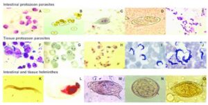
Types of Microscopic Parasites and How They’re Identified
Let’s look at a few major groups of parasites and how they appear under the microscope.
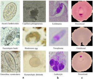
1. Protozoa (Single-celled organisms)
Plasmodium spp. (cause of malaria):
Seen in Giemsa-stained blood films.
Shows up as small ring-shaped or banana-shaped forms inside red blood cells.
Thick and thin blood smears are examined under oil immersion.
Used to determine parasite density and species (e.g. P. falciparum, P. vivax).
Entamoeba histolytica (cause of amoebic dysentery):
Found in stool samples.
Trophozoites (active stage) show a central karyosome and ingested red blood cells.
Cysts (dormant stage) have 1–4 nuclei.
Giardia lamblia:
Identified in stool samples using saline and iodine wet mounts.
Pear-shaped organism with two nuclei (like a “face”) and flagella.
2. Helminths (Worms)
Ascaris lumbricoides (roundworm):
Eggs are seen in stool samples.
Fertilised eggs are oval with thick shells; unfertilised ones are more irregular.
Hookworm:
Eggs are oval and thin-shelled with a clear space around the developing embryo.
Seen in stool using saline wet mount.
Enterobius vermicularis (pinworm):
Diagnosed using cellophane tape swab pressed around the anus early in the morning.
Eggs are flattened on one side and colourless.
3. Cestodes (Tapeworms)
Taenia spp.
Eggs have thick striated shells, sometimes visible in stool samples.
Proglottids (segments) passed in stool may also be examined to identify the species.
4. Trematodes (Flukes)
Schistosoma spp.
S. haematobium eggs (terminal spine) seen in urine samples.
S. mansoni eggs (lateral spine) seen in stool samples.
Staining and Preparation Methods
To enhance visibility and accuracy, several staining techniques and preparations are used:
Giemsa stain for blood parasites.
Iodine stain for stool protozoa.
Saline wet mount for observing motility.
Formalin-ether concentration to increase the number of parasites in stool for better viewing.
Imagine your younger cousin returns from school with a fever. At the hospital, a lab scientist takes a drop of blood and stains it on a slide. Under the microscope, they spot tiny rings inside red blood cells. That confirms it’s malaria caused by Plasmodium falciparum. That’s how simple and powerful microscopic identification can be.
Summary
- Microscopic identification is key to diagnosing parasitic infections.
- Blood, stool, and urine samples are examined using different stains and preparations.
- Each parasite has a unique appearance under the microscope that helps with diagnosis.
- Techniques like wet mount, Giemsa staining, and tape swabs are commonly used.
- This method is essential for managing parasitic diseases in everyday clinical settings.
Evaluation
- What stain is used to detect Plasmodium species in blood?
- How is Enterobius vermicularis usually diagnosed?
- What kind of sample is needed to detect Schistosoma haematobium?
- Name one protozoan and one helminth that can be seen in stool samples.
- Why is microscopic identification important in parasitic infections?
You’ve done an amazing job today! With every lesson, you’re building skills that could help diagnose and save lives in the future. Keep that energy high and remember, Afrilearn is always proud to walk beside you on your journey to greatness. See you at the next exciting class, champ!