Back to: MICROBIOLOGY 100 LEVEL
Welcome to class!
Hello brilliant mind! It’s another beautiful day to learn, and I’m super glad you’re here. You know how wearing different colours can make someone stand out or look sharper in pictures? That’s exactly what staining does in microbiology—it helps us see and understand microorganisms better under the microscope. Today, we’re going to learn about simple, differential, and special stains—what they are, why we use them, and how they help scientists study tiny life forms.
Simple, Differential, And Special Stains
What is Staining in Microbiology?
Most microorganisms, like bacteria, are nearly invisible under the microscope because they are colourless. To see them clearly, scientists apply stains (or dyes) that add colour to the organisms or parts of them. Think of it like using highlighter to focus attention on the most important part of a text.
Now, there are different types of stains depending on what you want to observe. Let’s break them down.
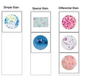
Simple Stains
This is the most basic type of staining. A simple stain uses only one type of dye to colour the entire microorganism. It makes it easier to see the size, shape, and arrangement of the cells.
For example, if you place some bacteria on a slide and add a blue dye like methylene blue, all the cells take up that colour and become visible.
This is like painting all the chairs in a room with one colour so you can easily count and describe them.
Differential Stains
As the name suggests, differential stains help tell the difference between types of microorganisms or between parts of a cell. They use two or more dyes and usually involve multiple steps.
The most common example is the Gram stain, which is used to divide bacteria into two groups:
Gram-positive bacteria appear purple.
Gram-negative bacteria appear pink or red.
This helps doctors and scientists choose the right antibiotics for an infection, because different bacteria respond to different treatments.
Another example is the acid-fast stain, which is used to detect Mycobacterium tuberculosis—the bacteria that causes TB.
Special Stains
Sometimes, scientists want to highlight only a particular part of the microorganism, like the capsule, flagella, or spores. That’s where special stains come in. These are specific techniques used to target just those parts.
For instance:
Capsule stain makes the slimy layer around some bacteria visible.
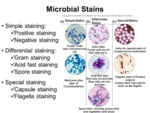
Endospore stain highlights the tough spores formed by some bacteria to survive harsh conditions.
Flagella stain shows the tail-like structures that help bacteria move.
These stains help scientists understand how dangerous or resilient a microorganism might be.
Summary
- Simple stains use one dye to show general features of the microorganism.
- Differential stains use more than one dye to distinguish between organisms or structures (e.g., Gram stain).
- Special stains focus on specific parts like spores, capsules, or flagella.
- Staining helps make invisible microbes visible and provides important information for diagnosis and research.
Evaluation
- What is the purpose of staining in microbiology?
- Name one dye used in simple staining.
- What colour does a Gram-positive bacterium appear after Gram staining?
- Which type of stain would you use to observe bacterial spores?
Today, you’ve learnt a powerful tool that microbiologists use every day in labs around the world. You’re becoming a real scientist, and Afrilearn is proud to be part of your journey. Keep going—we believe in you!