Back to: Botany 300 Level
Hello, my brilliant friend! I hope you’re having a fantastic day! Have you ever looked at a plant and wondered what it would look like up close, under a microscope? Just like humans have different tissues—like skin, muscles, and bones—plants also have specialised tissues that help them grow, transport nutrients, and survive in different environments. Today, we’re going to explore the microscopic world of plant tissues and learn how scientists examine them to understand how plants function!
Microscopic examination of plant tissues
Understanding the Microscopic Examination of Plant Tissues
Plants are made up of tiny cells that form different tissues. These tissues are responsible for support, transport, growth, and protection. To study them, scientists use a microscope, which magnifies plant tissues so that we can see the tiny structures inside them.
How to Examine Plant Tissues Under a Microscope
1. Collecting the Sample (Plant Tissue Preparation)
To examine a plant’s tissues, we first need a thin slice of the plant part we want to study. This could be a leaf, stem, or root. The sample must be thin so that light can pass through it.
2. Staining the Sample
Some plant tissues are almost transparent, so scientists use special dyes (stains) to highlight different parts of the cells. Common stains include:
Iodine solution – Stains starch in plant cells blue-black.
Safranin – Stains lignin in cell walls red.
Methylene blue – Stains cell nuclei blue.
3. Placing the Sample on a Microscope Slide
The stained sample is placed on a glass slide, covered with a thin glass coverslip, and examined under the light microscope.
4. Observing Different Plant Tissues
When we look through the microscope, we can see different types of plant tissues, each with unique structures and functions:
Types of Plant Tissues Seen Under a Microscope
Parenchyma Tissue
Structure: Thin-walled, loosely packed cells with large vacuoles.
Function: Stores food and water, helps in photosynthesis.
Example: Found in leaves, roots, and stems.
Collenchyma Tissue
Structure: Elongated cells with unevenly thickened walls.
Function: Provides flexible support to growing parts of the plant.
Example: Found in young stems and leaves (e.g., celery stalks).
Sclerenchyma Tissue
Structure: Thick-walled, dead cells with lignin.
Function: Provides strong support (e.g., in coconut shells, plant fibres).
Example: Found in seed coats, bark, and stems.
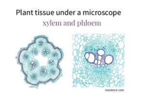
Xylem Tissue (Water Transport Tissue)
Structure: Long, tube-like cells with thick walls.
Function: Carries water and minerals from roots to leaves.
Example: Found in tree trunks and vascular bundles.
Phloem Tissue (Food Transport Tissue)
Structure: Tube-like cells called sieve elements with companion cells.
Function: Carries sugars and nutrients from leaves to other parts of the plant.
Example: Found in stems, roots, and leaves.
Epidermal Tissue (Protective Layer)
Structure: Thin layer of cells covering the plant surface.
Function: Protects the plant and prevents water loss.
Example: Found in leaves and stems, with stomata for gas exchange.
Why is Microscopic Examination of Plant Tissues Important?
Helps scientists understand how plants grow, transport nutrients, and survive.
Helps in plant breeding and genetic studies.
Helps identify plant diseases and nutrient deficiencies.
Used in agriculture and biotechnology to improve crop production.
A Simple Story to Understand This Concept
Imagine you are looking at a woven basket. From far, it looks like one object, but when you look closer, you see that it is made of tiny strands of fibres woven together. Similarly, a plant may look simple, but under a microscope, we can see the tiny cells and tissues working together to keep the plant alive!
Summary
Microscopes help us see the tiny structures of plant tissues.
Scientists use staining techniques to make plant cells more visible.
Different plant tissues (parenchyma, collenchyma, sclerenchyma, xylem, phloem, epidermis) have unique functions.
Studying plant tissues helps in scientific research, agriculture, and medicine.
Evaluation
- Why do scientists use a microscope to study plant tissues?
- What is the function of xylem and phloem tissues?
- How does staining help in plant tissue examination?
- What are the three types of simple plant tissues?
- Why is microscopic examination important in agriculture?
You are doing an amazing job! Now, the next time you see a leaf, remember that inside it, there are tiny, hardworking cells and tissues keeping the plant alive! Keep learning with Afrilearn, and I’ll see you in the next exciting lesson. Stay curious and keep growing!