Back to: HOME ECONOMICS JSS1
Welcome to class!
In today’s class, we will be talking about the eye. Enjoy the class!
The Eye

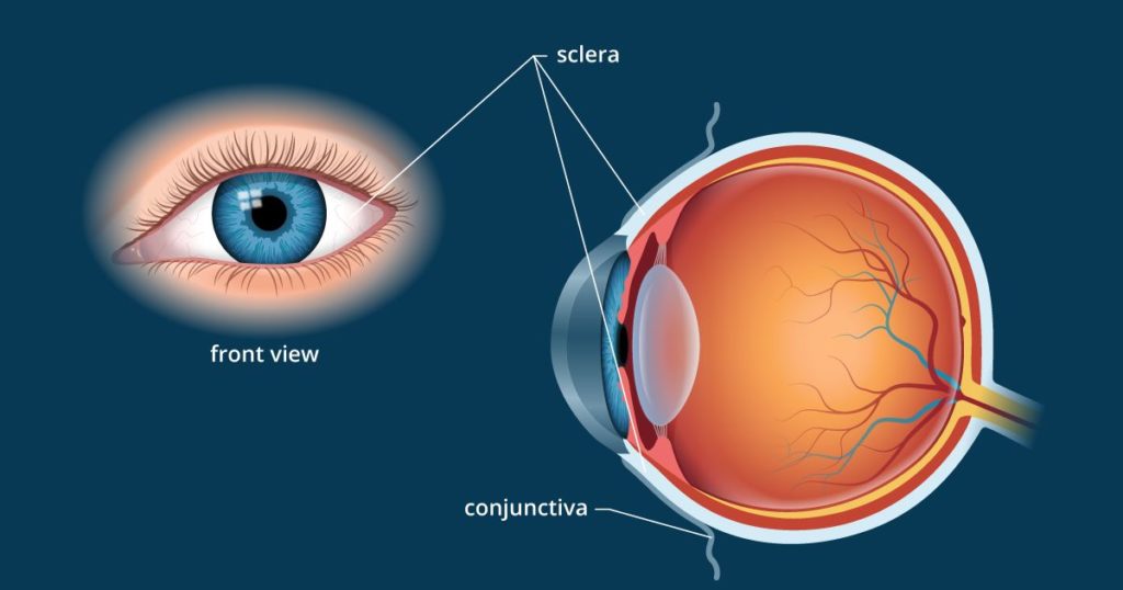
The eye is our organ of sight. The eye has several components which include but are not limited to the cornea, iris, pupil, lens, retina, macula, optic nerve, choroid and vitreous.
- Cornea: Clear front window of the eye that transmits and focuses light into the eye.
- Iris: Colored part of the eye that helps regulate the amount of light that enters
- Pupil: Dark aperture in the iris that determines how much light is let into the eye
- Lens: Transparent structure inside the eye that focuses light rays onto the retina
- Retina: Nerve layer that lines the back of the eye, senses light, and creates electrical impulses that travel through the optic nerve to the brain
- Macula: Small central area in the retina that contains special light-sensitive cells and allows us to see fine details clearly
- Optic nerve: Connects the eye to the brain and carries the electrical impulses formed by the retina to the visual cortex of the brain
- Vitreous: Clear, jelly-like substance that fills the middle of the eye

How the eye works
In several ways, the human eye works much like a digital camera:
- Light is focused primarily by the cornea— the clear front surface of the eye, which acts like a camera lens.
- The iris of the eye functions like the diaphragm of a camera, controlling the amount of light reaching the back of the eye by automatically adjusting the size of the pupil (aperture).
- The eye’s crystalline lens is located directly behind the pupil and further focuses light. Through a process called accommodation, this lens helps the eye automatically focus on near and approaching objects, like an auto-focus camera lens.
- Light focused by the cornea and crystalline lens (and limited by the iris and pupil) then reaches the retina — the light-sensitive inner lining of the back of the eye. The retina acts like an electronic image sensor of a digital camera, converting optical images into electronic signals. The optic nerve then transmits these signals to the visual cortex — the part of the brain that controls our sense of sight.
Layers of the eye
The fibrous tunic:
Also known as the tunica fibrosa oculi, is the outer layer of the eyeball consisting of the cornea and sclera. The sclera gives the eye most of its white colour. It consists of dense connective tissue filled with the protein collagen to both protect the inner components of the eye and maintain its shape.
The vascular tunic:
Also known as the tunica vascular oculior the “uvea”, is the middle vascularized layer which includes the iris, ciliary body, and choroid. The choroid contains blood vessels that supply the retinal cells with necessary oxygen and remove the waste products of respiration. The choroid gives the inner eye a dark colour, which prevents disruptive reflections within the eye.
The nervous tunic:
Also known as the tunica nervosa oculi, is the inner sensory layer which includes the retina. Contributing to vision, the retina contains the photosensitive rod and cone cells and associated neurons. To maximize vision and light absorption, the retina is a relatively smooth (but curved) layer. It has two points at which it is different; the fovea and optic disc.
In our next class, we will be talking about the Nose and the Ear. We hope you enjoyed the class.
Should you have any further question, feel free to ask in the comment section below and trust us to respond as soon as possible.

You are now doing a better job.
We’re glad you found it helpful😊 For even more class notes, engaging videos, and homework assistance, just download our Mobile App at https://play.google.com/store/apps/details?id=com.afrilearn. It’s packed with resources to help you succeed🌟