Back to: BIOLOGY SS3
Welcome to class!
In today’s class, we will be talking about sensory organs. Enjoy the class!
Sensory Organs

CONTENT
- Sensory receptors
- Skin as a sense organ
- The organ of sight, Structure of the eye
- Functions of the eye (Image formation and accommodation)
- Defects of the eye and their correction
Sensory receptors
All living organisms respond to changes in their environment (stimuli). These changes can be mechanical, electromagnetic, chemical or thermal. Though most cells in the bodies of organisms are sensitive to stimuli, certain cells specialize in detecting a particular type of stimulus, these are called sense cells or sensory receptors which are quite many in human bodies, monitoring the internal environment.
Sensory receptors sensitive to mechanical changes are called mechanoreceptors likewise thermoreceptors, chemoreceptors and photoreceptors are sensitive to heat, chemical and light respectively.
The sensory receptors change the detected stimulus into electrical impulses which when received by the brain are translated into pictures, sounds, smell, taste sensations. Structures containing sensory receptors are referred to as sense organs
A sense organ is defined as a group of specialized cells or tissues or which can receive, perceive or detect stimulus and transmit the information to the central nervous system. There are five types of sense organs in mammals; these include
- Skin detecting touch, pain, pressure and heat or cold.
- Eye detecting light (sense of sight)
- Ear detecting sound (sense of hearing and balancing)
- Nose detecting smell
- Tongue detecting taste
Evaluation
- What are sensory receptors
- What are sense organs? List major sense organs and their functions
Skin as a sense organ
In the mammalian skin, there are many sensory receptors for detecting several stimuli like touch, pressure, pain, cold and heat, unlike other sense organs which detect one type of stimulus each.
Generally, sensory receptors are not evenly distributed throughout the skin. Each type is more concentrated in a certain body region.
Sensory nerves ending sensitive to pressure (called pacinia corpuscles) are found deepest in the skin. Hence they need stronger stimulation. Those sensitive to touch (Meissner’s corpuscles) are largely distributed closest to the skin surface (in the epidermis) especially in hairless regions like tongue, fingers, lips, forehead etc. hence they need a gentle stimulation. In between pressure and touch, receptors are those detecting cold, heat and pain.
The organ of sight (Eye)
The eye is the organ of sight, spherical in shape and protected by ocular or optical structures like eye sockets, eyelids, eyelashes, tear or lacrimal glands and conjunctiva.
- The eye sockets house the eyes.
- Eyelids (upper and lower) protect the eye from foreign particles or mechanical injury.
- Tear or lacrimal glands, at the meeting point of the eyelids, secretes a salty fluid called tear which washes dust and destroys bacteria using its chemical substance called
- Eyelashes are rows of hairs on the eyelid which protect the eyeball from dust, excessive light and shield the eye against sweat and water.
- Conjunctiva is a thin transparent membrane lining the inside of the eyelids and protectively covers the cornea. The conjunctiva gets inflamed during infection (conjunctivitis).
Structure of the eye
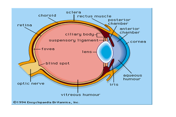
The wall of the eyeball consists of three layers namely (from outside inwards): sclera, choroid and retina
1. The sclera: the outermost white layer which gives shape to the eye and protects the inner part of the eye. The sclerotic layer bulges out in front of the eye to form the transparent cornea. The cornea admits light into the eye, brings the light to focus on the retina and protects the eye externally.
2. The choroid layer: This is highly vascularized and pigmented (black). This layer provides food and oxygen to the cells in the eye. The black pigment helps to absorb light rays and prevents light reflection. It consists of the ciliary muscles, iris, pupils, suspensory ligaments and the lens.
3. The retina: This is the part of the eye sensitive to light. It is also vascularized, pigmented and elastic. Light rays come to focus on the retina. Images formed on the retina are always real, inverted, and smaller than the real object. Two types of sensory cells (photoreceptors) found in retina are cones and rods.
- Cones are cells in the retina, which are sensitive to coloured visions and high light intensities. They contain a photochemical substance called iodopsin which is not easily bleached by high light intensities.
- Rods are more than the cones. They are sensitive to the colourless vision and low light intensities. A purple pigment-protein complex made from vitamin A called rhodopsin is found on the surface of rods. Rhodopsin is easily bleached when light falls on it.

- Yellow spot (Fovea centralis): This is the most sensitive part of the retina from where the fullest visual information is sent to the brain. It is the point where the image is focused.
- Blindspot: This is the point where the cells are not sensitive to light i.e no cones or rods The optic nerve goes out of the eye to the brain from the blind spot.
- Optic nerves: This nerve transmits sensory impulses to and from the brain.
- Aqueous humour: This is the transparent watery liquid which fills the space between the cornea and the lens. It is made up of solutions of protein, sugar, salt and water. This liquid refracts light rays onto the retina and helps to maintain the spherical shape of the eye.
- Vitreous humour: This is a wider, transparent, jelly-like liquid which fills the space between the lens and the retina. It is also a mixture of protein, sugar, salt and water. It carries out the same function as the aqueous humour.
Evaluation
- List five ocular structures and their functions
- State the functions of the following parts of the eye a) iris b) retina c) lens d) ciliary muscles
The function of the eye
The eye performs two major roles; Image formation and accommodation.
Image formation:
Light rays from any object pass enter the eye through the cornea, pass through the aqueous humour, lens and vitreous humour to the retina. These structures are all transparent and contribute to the refraction (bending) of the light rays thus enabling the rays to converge on the retina. The image of the object (real, inverted and smaller) is then formed on the retina. The stimulus of light reflected from the object is received by the rods or cones depending on the light intensity and are converted to an electrical impulse. The impulse is transmitted through the optic nerve to the optic lobe of the brain which correctly interprets the image. To form a sharp image of the object, all the light rays refracted meet at a particular point on the retina called yellow spot.
Accommodation:
This is the ability of the eye to focus near and distant objects on the retina i.e. the ability to see clearly through the adjustment of the focal length of the lens.
To see near objects
- The ciliary muscles contracts, making the suspensory ligaments relax their attention on the lens.
- The lens then becomes more convex in shape thus reducing the focal length of the lens to focus the image on the retina.
To see far objects
- The ciliary muscles relax, making the suspensory ligaments contract and pulling on the lens.
- The lens becomes flattened (elongated) increasing its focal length to focus the image on the retina.
Eye defects and corrections
An eye has defects when an image cannot be properly formed on the retina. The defects include;
- Short-sightedness (myopia): This is a defect in which a person sees nearby object clearly but distant ones appear blurred because the eyeball is longer than normal (from back to front). Therefore, light rays from a distant object are brought to focus in front of the retina. Correction: Using spectacles or glasses with suitable concave or diverging lens which diverge the light rays from a distant object before entering the eye so that the eye can bring the rays to a focus right on the retina.
- Long-sightedness (hypermetropia): This is the defect in which a person sees far object clearly but near ones appear blurred because the eyeball is shorter than normal. Therefore light rays from near objects are brought to a focus behind the retina. Correction: Using spectacles or glasses with suitable convex or converging lens which converge the light rays from the near object before entering the eye so that the eye can bring the rays to a focus right on the retina.
- Presbyopia: This is an eye defect resulting when the lens and the ciliary muscle lose their elasticity with advancing age. Therefore, light rays from nearby objects are not bent inward sufficiently and so are brought to a focus behind the retina. Correction: By the use of a converging lens.
- Astigmatism: This is caused by uneven cornea surface and can be corrected by using a lens with a compensating uneven surface.
- Cataract: This occurs mainly in old people in which eye lens becomes cloudy and can be corrected with a plastic lens or spectacles with a suitable lens.
- Night blindness: This is due to deficiency of vitamin A
- Conjunctivitis: Inflammation of the conjunctiva caused by the bacteria or irritants in the wind.
Evaluation
- Describe two major eye defects and their functions
- Describe the mechanism of image formation by the eye
In our next class, we will be talking about the Organ of Hearing, Smell and Taste. We hope you enjoyed the class.
Should you have any further question, feel free to ask in the comment section below and trust us to respond as soon as possible.

this is very helpful
We’re glad you found it helpful😊 For even more class notes, engaging videos, and homework assistance, just download our Mobile App at https://play.google.com/store/apps/details?id=com.afrilearn. It’s packed with resources to help you succeed🌟