Back to: AGRICULTURAL SCIENCE JSS2
Welcome to class!
Last class, we discussed Animal Diseases.
In today’s class, We will be diving deeper and sharing more about animal diseases. Enjoy the class!
ANIMAL DISEASES II
1. Tetanus
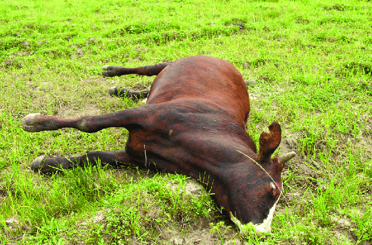
This is an infectious, non-febrile disease of animals and man, and is characterized by spasmodic tetany and hyperaesthesia. This disease is prevalent all over the world.
Transmission: Infection takes place by contamination of wounds. Deep puncture wounds provide favourable conditions for the spores to germinate, multiply and produce a toxin which is subsequently absorbed in the animal body. The micro-organism is present in soil and in animal faeces and is carried into the wound by a penetrating object. The organism is present in the intestine of normal animals, and under some undetermined conditions multiplies rapidly and produces the toxin in sufficient quantities to be absorbed and cause the disease.

Symptoms: The incubation period is generally 1-2 weeks but it may be as short as 3 days. Tetanus affects many species of domesticated animals but occurs particularly in horses and lambs; less frequently in adult sheep, goats, cattle, pigs, dog and cats; and rarely in poultry. The initial symptoms are mild stiffness and an unwillingness to move all the animals. More severe symptoms develop after 12-24 hours which are stiffness of limbs, neck, head, tail and twitching of muscles. The spasms develop in response to noise. In terminal stages ears are erect, nostrils dilated, nictitating membrane protruded. Mastication becomes very difficult because the mouth cannot be opened, hence the name lockjaw.
Treatment: The treatment is carried out by first injecting antitoxin then treating the wound. Penicillin parenterally is beneficial. Muscular relaxation is achieved by injection of relaxants. The animal should be kept in a dark room and fed with the help of a stomach tube.
Control: Proper hygiene and cleanliness at castration and other surgical procedures should be observed. Sheep should be given 2 injections based 3 weeks apart to develop a solid immunity.
2. Mastitis
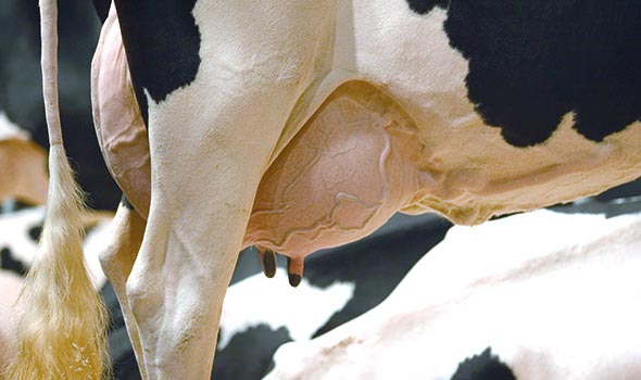
Mastitis, or inflammation of the mammary gland, is the most common and the most expensive disease of dairy cattle throughout most of the world. Although stress and physical injuries may cause inflammation of the gland, infection by invading bacteria or other microorganisms (fungi, yeasts and possibly viruses) is the primary cause of mastitis. Infections begin when microorganisms penetrate the teat canal and multiply in the mammary gland.

Treatment
- success depends on the nature of the aetiological agent involved, the severity of the disease and the extent of fibrosis.
- complete recovery with freedom from bacterial infection can be obtained in cases of recent infection and in those where fibrosis has taken place only to a small extent.
- such drugs as acriflavine, gramicidin and tyrothricin have now ceased to be in use, and have given place to the more effective drugs, such as sulphonamides, penicillin and streptomycin.
3. Footrot
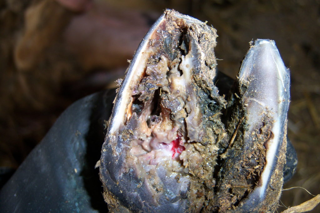
Footrot is a common cause of lameness in cattle and occurs most frequently when cattle on pasture are forced to walk through mud to obtain water and feed. However, it may occur among cattle in paddocks as well, under apparently excellent conditions. Footrot is caused when a cut or scratch in the skin allows the infection to penetrate between the claws or around the top of the hoof. Individual cases should be kept in a dry place and treated promptly with medication as directed by a veterinarian. If the disease becomes a herd problem a footbath containing a 5% solution of copper sulphate placed where cattle are forced to walk through it once or twice a day will help to reduce the number of new infections. In addition, drain mud holes and cement areas around the water troughs where cattle are likely to pick up the infection. Keep pens and areas where cattle gather as clean as possible. Proper nutrition regarding protein, minerals and vitamins will maximize hoof health.

- Ringworm
This is the most common infectious skin disease affecting beef cattle. It is caused by a fungus and is transmissible to man. Typically, the disease appears as crusty grey patches usually in the region of the head and neck and particularly around the eyes.
As a first step in controlling the disease, it is recommended that, whenever possible, affected animals should be segregated and their pens or stalls cleaned and disinfected. Clean cattle which have been in contact with the disease should be watched closely for the appearance of lesions and treated promptly. Proper nutrition, particularly high levels of vitamin a, copper and zinc while not a cure, will help to raise the resistance of the animal and in so doing offer some measure of control. Contact your vet and or feed store for products to treat this disease. Using a wormer like ivomec will kill lice and help prevent cattle from scratching causing skin damage and a place for the fungus to enter.
- Milk fever
Milk fever, also known as Parturient hypocalcaemia and parturient paresis, is a disease which has assumed considerable importance with the development of heavy milking cows. A decrease in the levels of ionized calcium in tissue fluids is basically the cause of the disease. In all adult cows, there is a fall in serum calcium level with the onset of lactation at calving. The disease usually occurs in 5 to 10-year-old cows and is chiefly caused by a sudden decrease in blood-calcium level, generally within 48 hours after calving.
Symptoms
- in classical cases, hypocalcaemia is the cause of clinical symptoms. Hypophosphataemia and variations in the concentration of serum-magnesium may play some subsidiary role.
- the clinical symptoms develop usually in one to three days after calving. They are characterized by loss of appetite, constipation and restlessness, but there is no rise in temperature.
- Calf scour
Calves may develop scours due to bacterial or viral infections. Scours is known as “calf scours” or neonatal calf diarrhoea. The primary causes of scours include Rotavirus, Coronavirus, Cryptosporidium parvum, Salmonella and Escherichia coli.
- Determine if treatment is required. Calves that are moving around in the pasture, with their tails up, probably do not need treatment. Check to see if the diarrhoea is yellow or white. If this is the case, treatment is probably not needed.
- Determine if the calf is looking listless. Calves that are lethargic or not participating much in the playful activities with other calves are a red flag to pay attention to. Calves that are also losing condition also cause for alarm.
- Check to see if the calf is dehydrated. You can check for dehydration by pulling on the calf’s neck skin. If the skin “tents” this is a sign of dehydration.
- Determine the calf’s body temperature. A normal body temperature ranges from 100.5 °f (38.1 °c) to 102.5 °f (39.2 °c). Anything outside of this range is a sign for treatment.
- Separate the sick calf or calves from the healthy herd. You’ll want to do this to avoid spreading the disease further.
- Administer fluids using your veterinarian-approved electrolyte solution. You may need to inject the fluids via iv or orally.
- Follow appropriate nursing care protocol using your vet’s guidelines. This may include providing shelter, feed and a warm place to sleep.
In our next class, we will be talking about Farm Animal’s Diseases. We do hope you enjoyed the class?
Should you have any further question, feel free to ask in the comment section below and trust us to respond as soon as possible.

I love this program 🥰🥰
good notes and how can I get it
it’s fun keep it up