Back to: BIOLOGY SS2
Welcome to SS2 Second Term!
We are eager to have you join us in class!!
In today’s class, We will be discussing the Tissue and Supporting System. We hope you enjoy the class!
TISSUE AND SUPPORTING SYSTEM
Contents
- Introduction
- Forms of Skeleton
- Types of Skeleton
- Functions of Skeleton
- Support in Vertebrates
- Axial and Appendicular Skeleton
- Supporting Tissues in Plants
INTRODUCTION
To carry out life processes, all organisms (plants and animals) need tissues. Tissues are a group of similar cells that carry out specific functions. Skeleton is the framework of the body which provides support, shape and protection to the soft tissues and organs in animals. It forms the central core of the human body and it is covered by muscles and blood vessels and skin.
FORMS OF SKELETAL MATERIALS
There are 3 forms of skeletal materials found in animals. These are
- Chitin
- Cartilage
- Bones
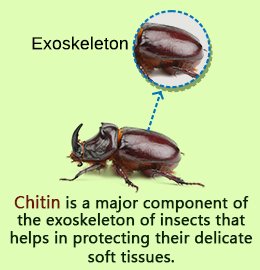
CHITIN
It is a tough non-living material present in arthropods (invertebrates). It acts as a hard outer covering to the animal and is made up of a series of plates covering or surrounding organisms. Chitin is very tough, light and flexible. However, it can be strengthened by impregnation with ‘tanned’ proteins and particularly in the aquatic crustaceans like crabs, by calcium carbonate.
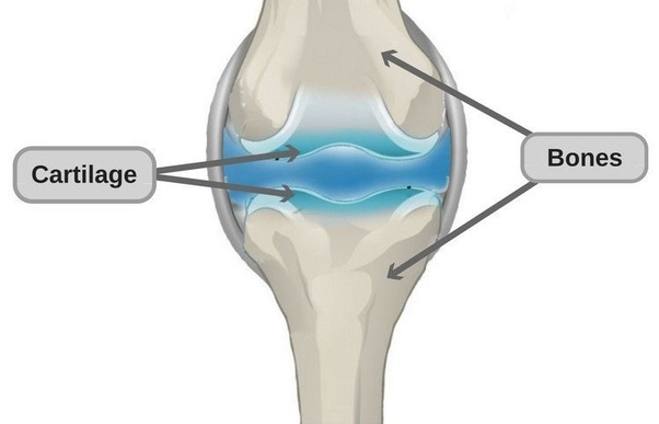
CARTILAGE
This is a tissue present in the skeleton of complex vertebrates. Cartilage consists of a hard matrix penetrated by numerous connective tissue fibres. The matrix is secreted by living cells called chondroblasts. These later become enclosed in spaces (lacunae) scattered throughout the matrix. In this condition, the cells are termed chondrocytes. It acts as a shock absorber in between bones during movement because it is tough and flexible with great tensile strength. It is found predominantly in mammals and cartilaginous fishes e.g. shark.
SELF EVALUATION
- What is a skeleton?
- (a) State two main components of the skeleton. (b) Differentiate between cartilage and chitin.
TYPES OF CARTILAGE
Cartilages are of three main types in mammals and they are
HYALINE CARTILAGE
This contains a dense meshwork is the most common type and can be found on the surface of moveable joint, trachea and bronchi (for ease of respiration) and also in protruding parts of the nose.
WHITE FIBROUS CARTILAGE
Tougher than the hyaline cartilage and can be found in the intervertebral disc of the vertebral column.
YELLOW ELASTIC CARTILAGE
Found in the external ear (pinna) and epiglottis (*cartilaginous flap covering the trachea active during food swallowing).
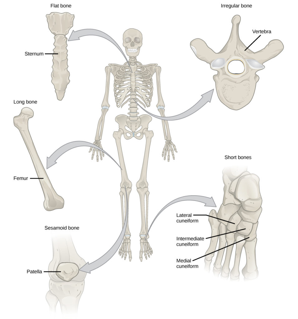
BONE
This is the major component of the skeletal system and it consists of living cells (osteocytes), protein fibres (collagen), and minerals such as calcium carbonate and calcium phosphate. These minerals (the non- living constituent) makes up two-thirds of a mass of bone. Hence, bone is strong and very rigid, unlike cartilage. Bones are highly vascularised.
The skeleton of a young vertebrate embryo is made up of cartilage. As the embryo grows bone cells (osteocyte) replaces cartilage cells. Hence, the cartilage tissue becomes hardened into the bone through the addition of minerals in a process called OSSIFICATION
Differences between Bones and Cartilage
| Bone | Cartilage | |
| 1 | Bones produce red and white blood cells | Cartilages do not |
| 2 | Made up of both living cells and dead cells | Made up of mainly living cells. |
| 3 | Bones are often rigid | Cartilages are often flexible |
| 4 | They are made up mainly of mineral substances such as calcium | Mineral substances are absent |
| 5 | Can never be replaced by cartilage | Can be replaced by bones |
| 6 | Flexible only in young ones | Cartilage both in young ones and adult is flexible. |
SELF EVALUATION
- With examples differentiate between hyaline and elastic cartilage.
- Distinguish between bone and cartilage.
TYPES OF SKELETON
The three main types of skeletons in animals are
- Hydrostatic skeleton: This is the type present in soft-bodied animals e.g. earthworm, sea anemones etc. Such animal use pressure to support itself. They also have a muscular body wall which is filled with fluid. The fluid presses against the muscular wall causing them to contract and exerting force against the fluid.
- Exoskeleton: This is the outer skeleton present in arthropods. It is secreted by the cells covering the body of the animals and the main component is chitin (non-living substance). The exoskeleton also supports animals against gravity and enables them to move about. Animals with these skeleton types periodically shed the old skeleton; grow rapidly in size when the new exoskeleton is still soft and extensible. The shedding process is called MOULTING or ECDYSIS.
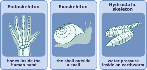
- Endoskeleton: This is an internal skeleton present in all vertebrates. Endoskeleton of vertebrates are composed mainly of bones and the bones grow steadily as the animal grows (hence no need for moulting). Bones of many sizes and shapes make up the endoskeleton of vertebrates. These bones are attached together as moveable joints by tough flexible fibres called ligaments hence the skeleton is flexible. Muscles are also attached to the bones usually by tendons to provide posture and bring about body movement.
FUNCTION OF SKELETON
- It supports the body of organisms.
- Skeleton acts as the framework of the body
- Protection of delicate organs e.g. heart, brain, etc.
- Used for locomotion through the limbs in action.
- An important component of respiration e.g. breathing involves active movement of the ribs.
- Production of blood via bone marrows.
SELF EVALUATION
- Mention the three types of skeleton.
- Differentiate between internal and external skeleton.
SUPPORT IN VERTEBRATES
The skeleton of vertebrates such as fish, frog, lizard, bird and man consists of bones and cartilages. It can be classified into two.
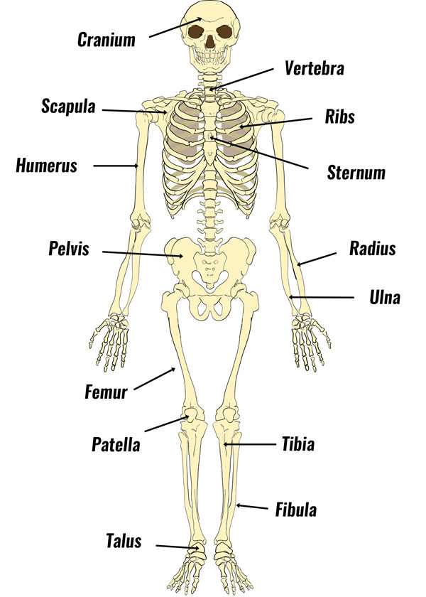
- AXIAL SKELETON- which consists of the skull, ribs, sternum and the vertebral column.
- APPENDICULAR SKELETON – it is made up of limb girdles (pectoral and pelvic girdles), and the limbs (the fore and hind limbs).
AXIAL SKELETON
The Skull
The Skull is made up of flat bones joined together by suture joint which has three parts: Cranium (brain-box), facial skeleton and the jaws; including maxilla (upper) and mandible.
Functions
- It protects the brain.
- Also protects the olfactory organ, eyes, middle and inner ear.
- Gives shape to the head.
- Bears the teeth.
The vertebral column
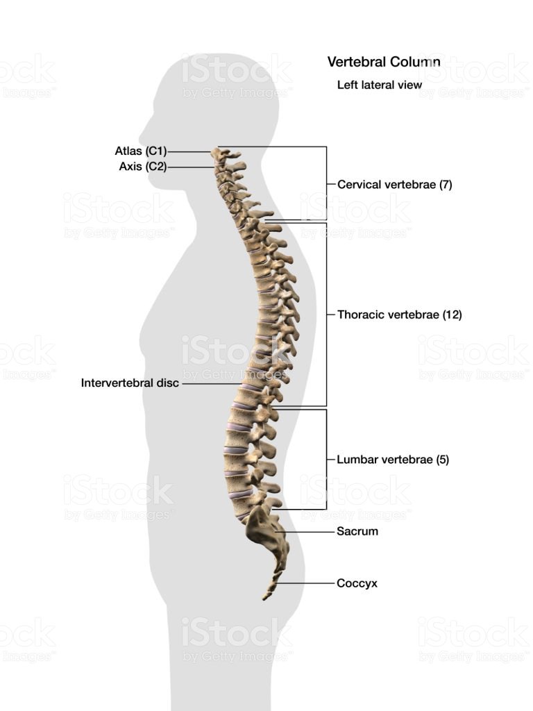
It forms the backbone, protecting the spinal cord. It is made up of 5 groups of bones called the vertebrate each of which is built on a similar basic pattern. The vertebrate is held together with strong ligament and comprehensible cartilage pads called into the intervertebral disc.
Types of Vertebrae and Location
| Vertebrae | Location | Rat | Rabbit | Cat | Cow | Humans |
| Cervical | Neck | 7 | 7 | 7 | 7 | 7 |
| Thoracic | Chest | 13 | 12-13 | 13 | 13 | 12 |
| Lumbar | Upper trunk | 6 | 6-7 | 7 | 6 | 5 |
| Sacral | Lower trunk | 4 | 4 | 3 | 5 | 5 |
| Caudal | Tail | 30 | 16 | 18-25 | 18-20 | 4 |
A TYPICAL VERTEBRAE
A typical vertebra has the following structural features
- A neural canal which is for the passage of the spinal cord.
- A neural spine which projects upward and backwards for the attachment of a muscle.
- Transverse processes for the attachment of muscles and ligaments.
- Centrum; solid bony pieces below the neural canal
- Zygapophyses are the particular surfaces for joining together of successive vertebrates.
This could be pre-zygapophysis (facing inward and upwards) or post-zygapophysis (facing outward and downwards
Cervical Vertebrae
The first cervical vertebra is called the atlas while the second is called the axis.
The Atlas It has a large neural canal, flat and broad transverse processes, short neural spine which could be absent at times. It also has a vertebrarterial canal for the passage of blood vessels. Centrum is absent.
SELF EVALUATION
- Give two classes of skeleton.
- List types of vertebrae.
Function of Atlas
Permits the nodding of the head.
The Axis
It has a broad and flat Centrum, a large and flat neural spine, reduce transverse processes and a vertebra arterial canal. It articulates with the atlas through odontoid process
Functions
- It permits the turning or twisting of the head.
- Forms pivot joint with the atlas.
Thoracic Vertebra
Have a long and prominent neural spine, a pair of short transverse processes, a large neural canal and neural arc and large cylindrical centrums. They also have particular surfaces for attachment of the ribs.
Function
- Aids attachment of ribs
- Assist in breathing
- Attachment of muscles at the shoulder and back
Lumbar Vertebrae
Each has large and flat transverse processes, broad and flat neural spine, large and thick centrums and well-developed zygapophyses. It has extra paired projections namely
- anapophysis
- metapophysis
Functions of Lumbar
- It provides attachment for abdominal muscles
- It bears a considerable weight of the body
Sacral Vertebrae
This fuse together to form a singular bony mass called the Sacrum. Each sacral vertebrate has a narrow neural canal, reduced neural spine and large centrums. The first differs from the remaining four by
- Having a pair of transverse processes which is large and wing-like while the others are attached to the muscles of the back.
- Presence of a small neural canal which generally becomes narrower in the lower four vertebrae.
FUNCTION
- Joins the pelvic girdle to provide support, rigidity and strength.
GENERAL SELF EVALUATION
- Describe the structural features of a typical vertebra
- Define ossification.
- What is moulting?
We have come to the end of this class. We do hope you enjoyed the class?
Should you have any further question, feel free to ask in the comment section below and trust us to respond as soon as possible.
In our next class, we will be talking about Tissue and Supporting System Cont’d. We are very much eager to meet you there.