Back to: BIOLOGY SS2
Welcome to Class !!
We are eager to have you join us !!
In today’s Biology class, We will continue learning about Tissues and Supporting System from our previous lesson. We hope you enjoy the class!
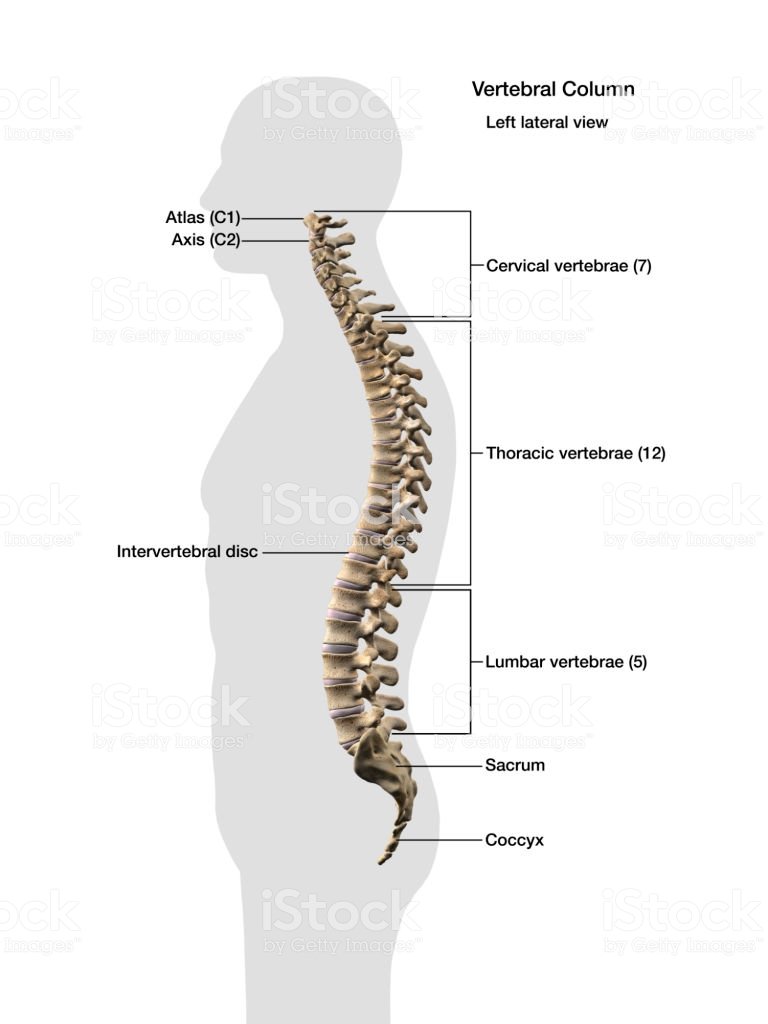
Caudal vertebrae
These are joined together to form a singular bony mass called the coccyx. Each has no neural spine, no neural canal and no transverse process. It appears as a solid rectangular mass of bone.
Functions
- Supports the tail
- Provides attachment for tail muscle
Evaluation
- List the structural features of a typical vertebrate
- Using the location, structural features and functions, differentiate between atlas and axis.
The Appendicular Skeleton
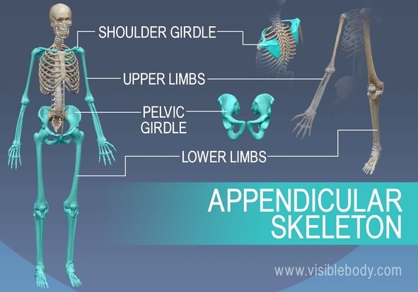
Pectoral/Shoulder girdle: found around the shoulder in man and it consists of two halves which are held
by muscles. Each half is made up of 3three bones
- Scapula
- Clavicle
- Coracoids
The scapula and coracoids are fixed together as the scapula is flat and triangular with a hollow called GLENOID CAVITY at its tip. This cavity articulates or joins with the head of the humerus to form the shoulder joint. The clavicle is a small rod of bone attached to a ligament joining the sternum to the scapula
Functions
- The pectoral girdle gives attachment to muscles and ligaments.
- It provides firm support to the forelimbs.
Pelvic girdle: found around the waist in man and it consists of two halves which are joined to each other ventrally and to the sacrum dorsally. Each half of the pelvic girdle is made up of 3threebones. They are
-
- Illium
- Ischium
- Pubis
These three bones form a depression (on their outer surface) called ACETABULUM which articulates with the head of the femur to form the hip joint.
Evaluation
- describe the limb-girdle found in the shoulder region of the human body
- Differentiate between pectoral and pelvic girdle.
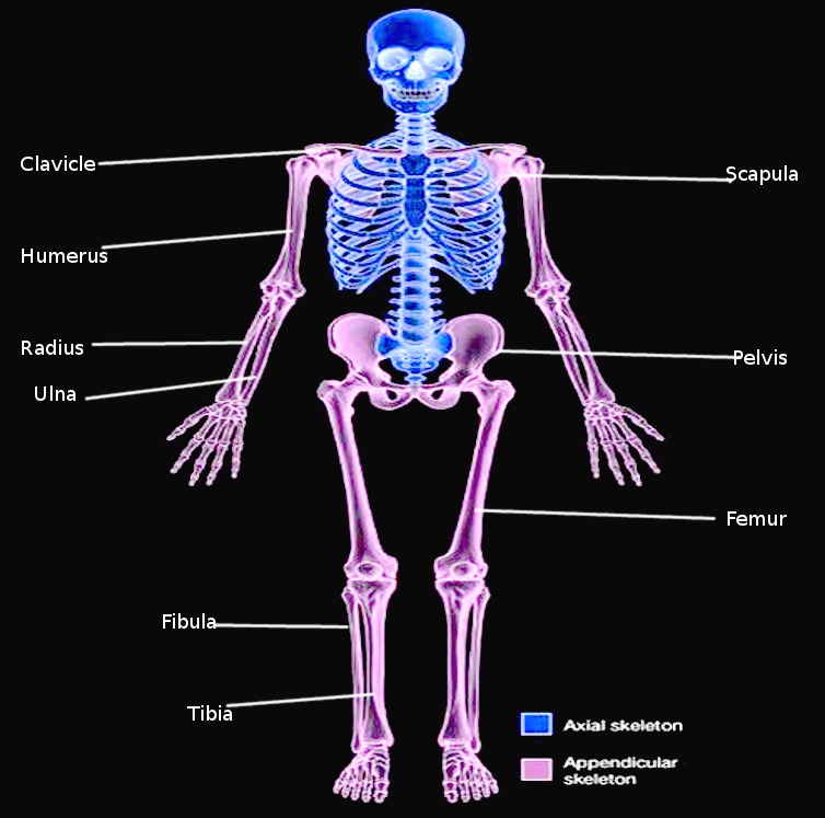
LIMBS
The limbs include the fore (upper) and the hind (lower) limbs. In most vertebrates, both limbs have the same basic plan i.e. each limb has a long bone followed by a pair of two long bones next to this is a set of small bones terminating with five digits.
The forelimbs– This is made up of an upper arm bone called humerus which joins with two other long bones at its lower end (radius and ulna) to form the elbow joints. Radius and ulna (the ulna is longer) are the bones of the forearm, next are the wrist bones called carpals which are small bones. These are followed by the digit bones called metacarpals which terminate in the phalanges (finger bones). In man, each digit has three phalanges except the thumb which has two phalanges.
The hind limbs-This is made up of thigh bones called femur (which is the largest and longest bone in the body). Its round upper end is the end that terminates at two rounded projections called condyles which forms the knee joint together with the tibia. A small flat bone called patella is found in front of the knee joint. Next to the femur are the tibia and fibula-Tibia is longer and larger. These are followed by bones of the ankle called tarsals. The lower limb terminates as at the digit bone metatarsals and each digit is made up of three phalanges
Evaluation
- on what formation plan are the upper and lower limb based
- Differentiate between the long bones of the arm and that of the thigh.
The ribs
These are long semi-circular rods which connect the thoracic vertebrates to the sternum. They are found in the chest region of the body. In man, they are 12 pairs
Function
- They form a cage protecting the lungs and the heart
- They assist in breathing.
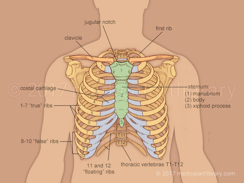
A TYPICAL RIB
A typical rib has a head, a neck and a body. The first seven ribs are connected directly to the sternum through costal cartilages. They are therefore called true ribs. The next five are called false ribs. The eighth to tenth ribs have a common articulation to the sternum, each one attached to the costal cartilage to the one above. The eleventh and twelfth pairs of ribs are called floating ribs because they have no connection to the sternum.
EVALUATION
- Describe the bone connecting the thoracic vertebra to the sternum.
- Classify the twelve types of ribs.
SUPPORTING TISSUES IN PLANTS
The needs for supporting tissues in a plant are for:
- definite shape;
- strength;
- rigidity;
- resistance against an external force such as wind and water.
Types of Supporting Tissues
- Parenchyma tissues
- Collenchyma tissues
- Sclerenchyma tissues
- Xylem tissues
- Phloem tissues
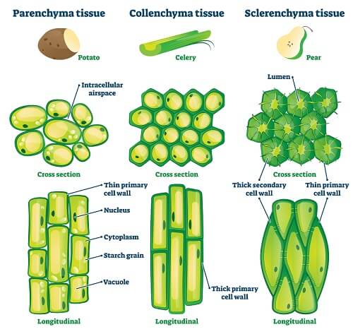
Parenchyma Tissues
They are made up living cells with cellulose and many air spaces between within them. This is the most common and abundant plant tissue.
Functions
- It gives firms and turgidity to the stems of hibiscus
- stores food and water
- takes part in food synthesis in leaf mesophyll
Collenchyma Tissues
Made up of living cells which are elongated and thickened at the corners.
Functions
- Provides strength and support in a young growing plant.
- Gives flexibility and resilience to plant.
Sclerenchyma Tissues
They are made up of thick cells containing cellulose and lignin. The tissues are rich in fibres.
Functions
- gives flexibility to plant
- provides strength, rigidity, hardness and support to plant
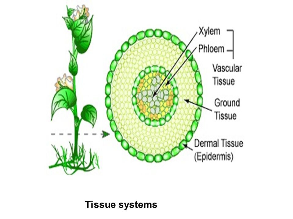
Xylem
Xylem tissues are found in vascular tissues of stems, roots and leaves
Functions
- provides support strength and shape to the plant
- Helps to conduct water and mineral salt from the roots to leaves.
Phloem Tissues
Also located in the vascular bundles of all plants in their roots, stems and leaves
Functions
- Conduction of manufactured food from the site of production to the site of consumption and storage.
- Assist to provide support to the entire plant.
GENERAL EVALUATION
- Describe the structural features of a typical vertebra
- Define ossification.
- What is moulting?
- State four reasons for the presence of supporting tissues in plant
- List supporting tissues found in plant and state their functions.
Reading Assignment
College Biology by Idodo Umeh. Chapter14, page 251-259
WEEKEND ASSIGNMENT
SECTION A
- …………. is a non-living skeletal material (a) chondroblasts B. osteocyte C. elastic cartilage D. chitin
- The articulating surface for joining together successive vertebrates is called (a) neural spine B. zygapophyses C. transverse processes D. neural canal
- The canal for the passage of blood vessels in vertebrae is called. (a) Neural canal B. cervical canal C. vertebral canal D. zygaphosis
- Endoskeleton is present in the following animals except (a) dog B. snake C. shark D. lizard
- The most abundant supporting tissue in plants is (a) sclerenchyma B. parenchyma C. xylem D. phloem
SECTION B
- (a) What is ecdysis? (b) Mention two animals in which it occurs
- Differentiate between (a) Bone and cartilage (b) Atlas and axis vertebrae
We have come to the end of this class. We do hope you enjoyed the class?
Should you have any further question, feel free to ask in the comment section below and trust us to respond as soon as possible.
In our next class, we will be learning about Mammalian Teeth. We are very much eager to meet you there.